253,50 €*
Versandkostenfrei per Post / DHL
Aktuell nicht verfügbar
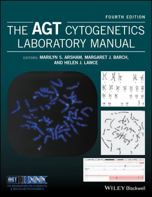
Zellgenetiker greifen auf eine ganze Reihe von Verfahren zurück, um Chromosomen und/oder eine Zielregion eines bestimmten Chromosom in der Meta- oder Interphase zu untersuchen. Zu den Tools gehören Routineanalysen von Chromosomen (G-Banding), spezielle Stains für bestimmte Chromosomenstrukturen und molekulare Sonden, z. B. im Bereich der Fluoreszenz-in-situ-Hybridisierung (FISH) sowie der Chromosomenanalyse auf Microarray-Basis. Zum Einsatz kommen eine Vielzahl von Methoden, um eine zu untersuchenden Region hervorzuheben, der so klein ist, wie eine einzige, spezifische Gensequenz.
Zellgenetiker greifen auf eine ganze Reihe von Verfahren zurück, um Chromosomen und/oder eine Zielregion eines bestimmten Chromosom in der Meta- oder Interphase zu untersuchen. Zu den Tools gehören Routineanalysen von Chromosomen (G-Banding), spezielle Stains für bestimmte Chromosomenstrukturen und molekulare Sonden, z. B. im Bereich der Fluoreszenz-in-situ-Hybridisierung (FISH) sowie der Chromosomenanalyse auf Microarray-Basis. Zum Einsatz kommen eine Vielzahl von Methoden, um eine zu untersuchenden Region hervorzuheben, der so klein ist, wie eine einzige, spezifische Gensequenz.
About the Editors
Marilyn S. Arsham, (retired) Cytogenetic Technologist II, Western Connecticut Health Network, Danbury Hospital campus, Danbury, Connecticut, USA
Margaret J. Barch, (formerly) Frank F Yen Cytogenetics Laboratory, Weisskopf Child Evaluation Center, University of Louisville, USA
Helen J. Lawce, Clinical Cytogenetics, Oregon Health & Science University Knight Diagnostics Laboratory, USA
Preface xxix
Acknowledgments xxxi
1 The cell and cell division 1
Margaret J. Barch and Helen J. Lawce
1.1 The cell 1
1.2 The cell cycle 14
1.3 Recombinant DNA techniques 19
1.4 The human genome 21
References 22
2 Cytogenetics: an overview 25
Helen J. Lawce and Michael G. Brown
2.1 Introduction 25
2.2 History of human cytogenetics 25
2.3 Cytogenetics methods 29
2.4 Slide-making 49
2.5 Chromosome staining 58
2.6 Chromosome microscopy/analysis 59
2.7 Laboratory procedure manual 69
References 70
Contributed protocols 75
Protocol 2.1 Slide-making 75
Protocol 2.2 Slide-making 76
Protocol 2.3 Making wet slides for chromosome analysis 78
Protocol 2.4 Slide-making 82
Protocol 2.5 Slide preparation 82
Protocol 2.6 Slide preparation procedure 84
3 Peripheral blood cytogenetic methods 87
Helen J. Lawce and Michael G. Brown
3.1 Using peripheral blood for cytogenetic analysis 87
3.2 Special uses of peripheral blood cultures 88
3.3 Peripheral blood constituents 89
3.4 Specimen handling 91
3.5 Cell culture equipment and supplies 93
3.6 Harvesting peripheral blood cultures 95
3.7 Chromosome analysis of peripheral blood 95
3.8 Storage of fixed specimens 95
Acknowledgments 95
References 95
Contributed protocols 98
Protocol 3.1 Blood culture and harvest procedure 98
Protocol 3.2 High-resolution peripheral blood method 100
Protocol 3.3 Constitutional cytogenetic studies on peripheral blood 108
Protocol 3.4 Blood culture and harvest procedure for microarray confirmation studies 115
4 General cell culture principles and fibroblast culture 119
Debra F. Saxe, Kristin M. May and Jean H. Priest
4.1 Definitions of a culture 119
4.2 Basic considerations in cell culture 121
4.3 Fibroblast culture 128
4.4 Lymphoblastoid cell lines 132
Glossary 132
Reference 133
Additional readings 133
Contributed protocols section 134
Protocol 4.1 Solid tissue collection for establishing cultures 134
Protocol 4.2 Solid tissue transport and sendout media 135
Protocol 4.3 Tissue culture reagents 138
Protocol 4.4 Phosphate buffer solution deficient in Ca2+ and Mg2+ 141
Protocol 4.5 Solid tissue and fibroblast culture setup 141
Protocol 4.6 Solid tissue setup and processing 142
Protocol 4.7 Flask and coverslip setup for POC/fibroblast cultures 145
Protocol 4.8 Coverslip setup for solid tissue biopsy specimens 147
Protocol 4.9 Solid tissue (fibroblast) culturing and harvesting 150
Protocol 4.10 Fibroblast culture maintenance: media feeding and changing 154
Protocol 4.11 Routine subculture of fibroblast cultures 155
Protocol 4.12 Manual harvest for flasks 157
Protocol 4.13 Treated media for contamination 158
Protocol 4.14 Fungizone-mycostatin solution for treatment of fungus/yeast contaminated cultures 158
Protocol 4.15 Mycoplasma testing 159
Protocol 4.16 Plating efficiency of serum 160
Protocol 4.17 Routine replication plating for human diploid cells 160
Protocol 4.18 Cell counting chamber method 161
Protocol 4.19 Cell viability by dye exclusion 161
Protocol 4.20 Mitotic index 161
Protocol 4.21 Growth rate-estimation of mean population doubling time during logarithmic growth 162
Protocol 4.22 Maintenance of fibroblast cultures as non-mitotic population 163
Protocol 4.23 Synchronization at S-phase with BrdU 163
Protocol 4.24 Making direct FISH preparations from abortus tissue 164
Protocol 4.25 Cryopreservation 165
Protocol 4.26 Cryopreservation with Nalgene cryogenic container 166
Protocol 4.27 Lymphoblastoid lines 167
Protocol 4.28 Freezing tissue cultures (cryopreservation) 171
5 Prenatal chromosome diagnosis 173
Kristin M. May, Debra F. Saxe and Jean H. Priest
5.1 Introduction 173
5.2 Amniotic fluid 173
5.3 Culture of amniotic fluid 175
5.4 Analysis of amniotic fluid 178
5.5 Chorionic villus sampling 180
5.6 Analysis of chorionic villi 184
References 186
Contributed protocols section 188
Protocol 5.1 Amniotic fluid culture setup and routine maintenance 188
Protocol 5.2 Coverslip (in situ) harvest procedure for chromosome preparations from amniotic fluid, CVS, or tissues (manual method) 191
Protocol 5.3 Harvest of flask amniocyte cultures 193
Protocol 5.4 Amniotic fluid culturing, subculturing, and harvesting (flask method) 195
Protocol 5.5 Criteria for interpreting mosaic amniotic fluid cultures 198
Protocol 5.6 Chorionic villi sampling - setup, direct harvest, and culture 199
Protocol 5.7 Chorionic villus sampling 204
Protocol 5.8 G-Banding with Leishman's stain (GTL) 208
Protocol 5.9 Cystic hygroma fluid protocol 209
6 Chromosome stains 213
Helen J. Lawce
6.1 Introduction 213
6.2 Chromosome banding methods 220
6.3 5-bromo-2'-deoxyuridine methodologies 246
6.4 T-banding/CT-banding 252
6.5 Antibody banding and restriction endonuclease banding 252
6.6 Destaining slides 252
6.7 FISH DAPI bands 252
6.8 Sequential staining 253
Acknowledgments 253
References 253
Contributed protocols section 266
Protocol 6.1 Conventional Giemsa staining (unbanded) 266
Protocol 6.2 Leishman's stain 266
Protocol 6.3 Quinacrine mustard chromosome staining (Q-bands) 266
Protocol 6.4 C-banding 268
Protocol 6.5 C-banding 270
Protocol 6.6 C-banding 271
Protocol 6.7 C-banding of blood slides 272
Protocol 6.8 Giemsa-11 staining technique 274
Protocol 6.9 Distamycin A/DAPI staining 275
Protocol 6.10 Chromomycin/methyl green and chromomycin/distamycin fluorescent R-banding method 277
Protocol 6.11 Bone marrow and cancer blood G-banding 278
Protocol 6.12 Trypsin G-banding 280
Protocol 6.13 Giemsa-trypsin banding with Wright stain (GTW) for suspension culture slides and in situ culture coverslips 281
Protocol 6.14 G-banding blood lymphocyte slides 284
Protocol 6.15 Cd staining 285
Protocol 6.16 CREST/CENP antibody staining 286
Protocol 6.17 AgNOR (silver staining) 287
Protocol 6.18 Sister chromatid exchange blood culture and staining 289
Protocol 6.19 Sister chromatid exchange fibroblast culture and staining 291
Protocol 6.20 T-banding by thermal denaturation 294
Protocol 6.21 CT-banding 295
Protocol 6.22 Lymphocyte culture and staining procedures for late replication analysis 295
Protocol 6.23 Destaining and sequential staining of slides 298
Protocol 6.24 Restaining permanently mounted slides 299
7 Human chromosomes: identification and variations 301
Helen J. Lawce and Luke Boyd
7.1 Understanding the basics 301
7.2 Description of human chromosome shapes 302
7.3 Determination of G-banded chromosome resolution 355
Acknowledgments 356
Glossary 356
References 357
8 ISCN: the universal language of cytogenetics 359
Marilyn S. Arsham and Lisa G. Shaffer
8.1 Introduction 359
8.2 Language 359
8.3 Karyotype 364
8.4 Numerical events 378
8.5 Structural events 380
8.6 Derivative chromosomes (der) 394
8.7 Symbols of uncertainty 397
8.8 Random versus reportable 403
8.9 Multiple cell lines and clones 404
8.10 Fluorescence in situ hybridization 408
8.11 Microarray (arr) and region-specific assay (rsa) 420
8.12 Conclusion 422
Acknowledgments 422
Addendum for ISCN 2016 updates 426
References 426
9 Constitutional chromosome abnormalities 429
Kathleen Kaiser-Rogers
9.1 Numerical abnormalities 429
9.2 Structural rearrangements 444
References 472
10 Genomic imprinting 481
R. Ellen Magenis
10.1 Introduction 481
10.2 Human genomic disease and imprinting 488
10.3 Germ cell tumors - UPD and imprinting 493
Glossary 494
References 496
11 Cytogenetic analysis of hematologic malignant diseases 499
Nyla A. Heerema
11.1 Introduction 499
11.2 Myeloid leukemias 508
11.3 Myelodysplastic syndromes 514
11.4 Myeloproliferative neoplasms 515
11.5 B- and T-cell lymphoid neoplasms 517
11.6 Lymphomas 522
11.7 Laboratory practices 525
Acknowledgments 533
Glossary of hematopoietic malignancies 533
References 535
Contributed protocols section 553
Protocol 11.1 Cancer cytogenetics procedure 553
Protocol 11.2 Bone marrow/leukemic peripheral blood setup and harvest procedure 558
Protocol 11.3 Bone marrow and leukemic blood culture and harvest procedure using DSP30 CPG oligonucleotide/interleukin-2 for B-cell mitogenic stimulation 560
Protocol 11.4 Culture of CpG-stimulated peripheral blood and bone marrow in chronic lymphocytic leukemia 562
Protocol 11.5 Plasma cell separation and harvest procedure for FISH analysis 567
Protocol 11.6 Plasma cell separation and harvest procedure for FISH 569
Protocol 11.7 Bone marrow GTG-banding 571
Protocol 11.8 GTW banding procedure (G-bands by trypsin using Wright stain) 573
12 Cytogenetic methods and findings in human solid tumors 577
Marilu Nelson
12.1 Introduction 577
12.2 Processing tumor specimens 579
12.3 Recurrent cytogenetic abnormalities 592
12.4 Molecular genetic and cytogenetic techniques 608
12.5 Conclusion 612
Glossary 612
References 613
Contributed protocol section 631
Protocol 12.1 Solid tumor cell culture and harvest 631
Protocol 12.2 Solid tumor cell culture and harvest 637
Protocol 12.3 Solid tumor culture 643
Protocol 12.4 Solid tumor harvest: monolayer and flask methods 644
Protocol 12.5 Solid tumor culturing and harvesting 646
13 Chromosome instability syndromes 653
Yassmine Akkari
13.1 Introduction 653
13.2 Fanconi anemia 656
13.3 Bloom syndrome 658
13.4 Ataxia-telangiectasia 658
13.5 Nijmegen breakage syndrome 659
13.6 Immunodeficiency, centromeric instability, and facial anomalies syndrome 660
13.7 Roberts syndrome 661
13.8 Werner syndrome 661
13.9 Rothmund-Thomson syndrome 662
13.10 Proficiency testing 662
Glossary 662
References 667
Contributed protocol section 671
Protocol 13.1 Fanconi anemia chromosome breakage procedure for whole blood 671
Protocol 13.2 Supplemental procedure; Ficoll separation of whole blood 675
Protocol 13.3 Fanconi anemia fibroblast set up, culture, subculture, and harvest procedure 676
Protocol 13.4 Fanconi anemia chromosome breakage analysis policy 681
Protocol 13.5 Table for breakage studies result...
| Erscheinungsjahr: | 2017 |
|---|---|
| Fachbereich: | Gentechnologie |
| Genre: | Biologie |
| Rubrik: | Naturwissenschaften & Technik |
| Medium: | Buch |
| Seiten: | 1168 |
| Inhalt: | 1168 S. |
| ISBN-13: | 9781119061229 |
| ISBN-10: | 1119061229 |
| Sprache: | Englisch |
| Herstellernummer: | 1W119061220 |
| Einband: | Gebunden |
| Autor: |
Arsham, Marilyn
Lawce, Helen Barch, Margaret |
| Redaktion: |
Arsham, Marilyn S
Barch, Margaret J Lawce, Helen J |
| Herausgeber: | Marilyn S Arsham/Margaret J Barch/Helen J Lawce |
| Auflage: | 4th edition |
| Hersteller: | John Wiley & Sons |
| Maße: | 287 x 220 x 53 mm |
| Von/Mit: | Marilyn S Arsham (u. a.) |
| Erscheinungsdatum: | 24.04.2017 |
| Gewicht: | 3,203 kg |

